Figure 3.

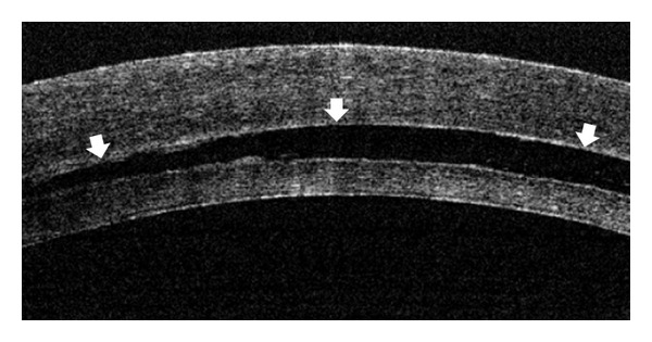
An OCT image of the eye of patient 2 at the end of the first DSEAK surgery. Interface fluid was observed between the host cornea and the donor graft (arrows).

An OCT image of the eye of patient 2 at the end of the first DSEAK surgery. Interface fluid was observed between the host cornea and the donor graft (arrows).