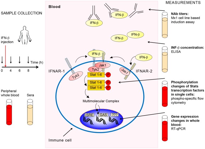
Figure 1. Schematic representation of workflow.
The workflow diagram shows times and type of sample collection before and after IFN-β administration and sample processing and analysis. Concentrations of NAbs (against IFN-β) and IFN-β were measured in sera. mRNA used for gene expression measurements was extracted from whole blood. For signaling pathway analysis by phosphoflow, immune cell subtypes were identified and phosphorylation levels of various Stat proteins quantified in single cells. If present in sera, NAbs prevent the initiation of the IFN-β signal at the cell surface. In the absence of NAbs IFN-β binds to its cognate receptor and forms an activated receptor complex with associated kinases that phosphorylate Stat proteins at specific residues. The Jak/Stat pathway is the major signaling pathway activated by IFN-β.

