
Fig. 3.
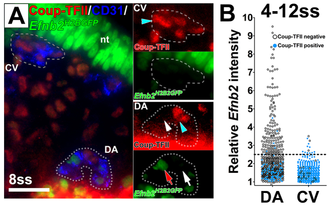
A heterogeneous population of endothelial cells in the dorsal aorta and cardinal vein expressed either the arterial marker Efnb2 or the venous marker Coup-TFII. (A) Cross-section of an Efnb2H2BGFP/+ embryo stained for CD31 (blue) and Coup-TFII (red). Insets show magnified views of the DA and CV. Coup-TFII (blue arrowheads) and Efnb2 (red arrow) were not detected in the same EC in either the DA or the CV. White arrow indicates Efnb2-negative ECs and white arrowhead Coup-TFII-negative ECs. (B) Quantification of Efnb2 intensity in Coup-TFII-stained Efnb2H2BGFP/+ embryos at 4-12 ss. Relative Efnb2 intensity above 2.5 was considered positive (dotted line). Blue circles indicate Coup-TFII-positive ECs and black circles indicate Coup-TFII-negative ECs. The Efnb2 intensity was below 2.5 for most Coup-TFII-positive ECs. nt: neural tube. CV, cardinal vein; DA, dorsal aorta; nt, neural tube. Scale bar: 25 μm.