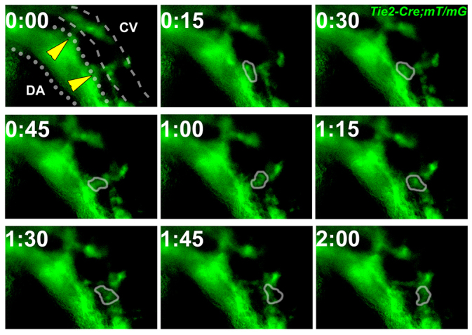
Fig. 4.

Endothelial cells moved from the dorsal aorta to the cardinal vein. Time-lapse imaging of EC movement in a Tie2-Cre;mT/mG embryo at 10 ss. Membrane-targeted GFP (green) is expressed by cells of the Tie2 lineage. During a 2-hour time frame, GFP-positive cell(s) with EC morphology (circled) moved from the DA (dotted line) to the CV (dashed line). Yellow arrowheads indicate connections between the DA and CV. CV, cardinal vein; DA, dorsal aorta.
