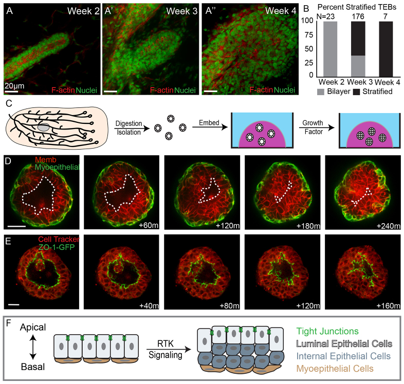
Fig. 1.
Mammary stratification generates an internal population of luminal epithelial cells lacking tight junctions. (A-A′) Terminal end buds from 2- to 4-week-old mice were stained for F-actin (red) and nuclei (green). (B) Quantification of bilayered (gray) and stratified (black) end buds at 2, 3 and 4 weeks postnatal. (C) A schematic depicting organotypic culture. (D) Still images from a movie of an organoid undergoing stratification. Luminal epithelial cells are labeled red with a membrane localized tdTomato and myoepithelial cells are labeled green with a genetically encoded green fluorescent protein (GFP). The dashed line highlights the boundary of the luminal space. (E) Still images of an organoid undergoing stratification with ZO-1-GFP marking tight junctions and Cell Tracker Red staining the cytosol. (F) A cartoon depiction of mammary epithelial stratification showing the generation of an internal luminal epithelial cell population lacking tight junctions. Scale bars: 20 μm.

