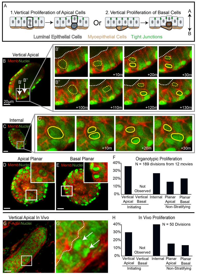
Fig. 2.
Vertical apical cell divisions initiated mammary stratification. (A) A schematic depicting the alternatives of stratification initiation by vertical apical versus vertical basal proliferation. (B-E) Frames from movies of organoids expressing nuclear (green) and membrane (red) markers were collected to visualize proliferation in real time. (B) An image of an organoid before stratification. The arrows highlight apical cells that undergo vertical proliferation in B′ and B′. (B′,B′) Movie frames showing two vertical apical cell divisions, each producing one apical mother cell and one internal daughter cell. Yellow dashed lines show apical mother cells, solid yellow lines highlight internal daughter cells and dashed white lines represent the border of the lumen. (C) An organoid that is partially stratified with inset on an internal cell that divides in C′. (C′) Frames showing division of the internal cell highlighted in C; solid yellow lines outline the dividing nuclei. (D,E) An image of planar proliferation of apical and basal cells, respectively, insets on dividing cells. (F) Quantification of the types of proliferation observed in organotypic culture. (G) A terminal end bud from 3-week-old mouse stained for F-actin (red) and nuclei (green). The inset highlights a vertical apical division. (G′) An enlarged image of the inset from G; the arrows point to dividing nuclei and the dashed white line shows the border of the lumen. (H) Quantification of the types of proliferation observed in vitro. Scale bars: 20 μm.

