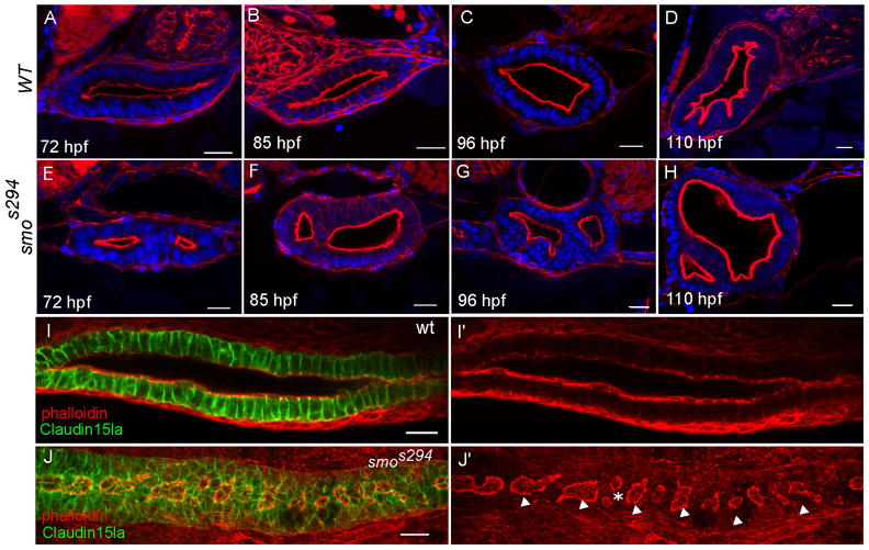
Fig. 5.
smos294 mutants exhibit a lumen fusion defect. (A-H) Confocal cross sections of wild-type (top) and smos294 (bottom) intestines at 72 hpf, 85 hpf, 96 hpf and 110 hpf. Phalloidin (red). (I,J) Confocal whole-mount image of wild-type and smos294 embryos expressing TgBAC(cldn15la-GFP) to highlight the cellular and luminal architecture of the intestine at 72 hpf. (I) Wild-type intestine, (J) smos294 intestine. Arrowheads, lumens; asterisk, adjacent lumens. Scale bars: 20 μm.

