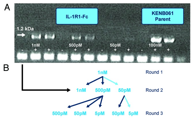
Figure 2. Selection strategy used for phage and ribosome display to generate potent anti-IL-1RI Antibodies. (A) Gel photograph of cDNA outputs from Round 2 of Ribosome Display Selections with antigen concentrations at 1 nM, 500 pM and 50 pM. (B) Selection pathway (high-lighted in light blue) followed to generate high affinity IL-1RI variants using phage and ribosome display.

An official website of the United States government
Here's how you know
Official websites use .gov
A
.gov website belongs to an official
government organization in the United States.
Secure .gov websites use HTTPS
A lock (
) or https:// means you've safely
connected to the .gov website. Share sensitive
information only on official, secure websites.
