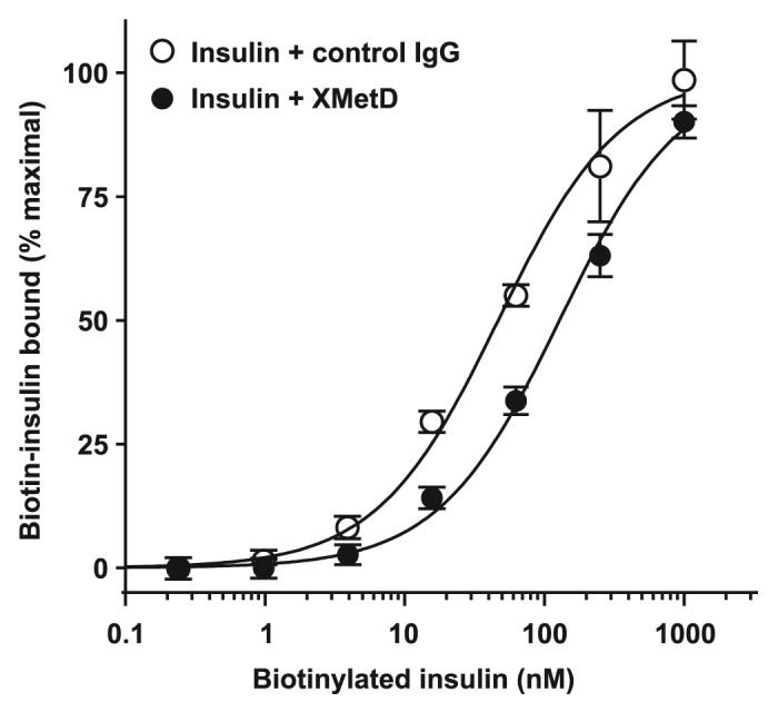
Figure 3. Insulin binding to the hINSR by FACS. CHO-hINSR cells (B isoform) were preincubated for 10 min at 4 ° with 1 μM of either XMetD or control IgG followed by a 30 min incubation with increasing concentrations of biotinylated insulin. Biotinylated insulin binding to the INSR was measured by flow cytometry (FACS). Mean ± SD of triplicate determinations are shown.
