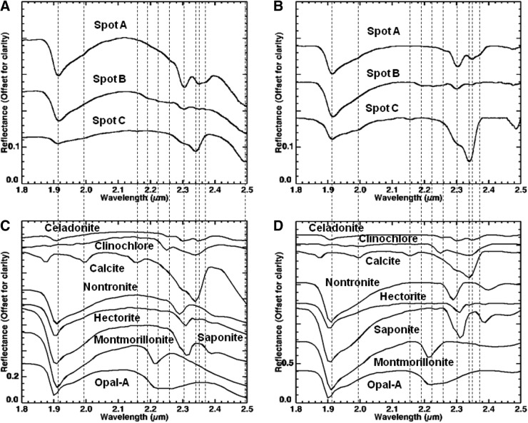
FIG. 16.
Close-up of spectral region 1.8–2.5 μm from Fig. 15, where VSWIR spectra (A and B) collected from spots (A–C) on sample 14 (Fig. 14) with laboratory spectrometer are compared with representative matching library spectra (C and D). Vertical lines correspond to features at 1.91, 1.99, 2.16, 2.19, 2.22, 2.26, 2.3, 2.34, 2.35, 2.37, and 2.5 μm. See text for details on features and discussion. Spectra on the left are normal, while spectra on the right are continuum-removed. All spectra are offset for clarity. For details on library spectra used, see Appendix A.

