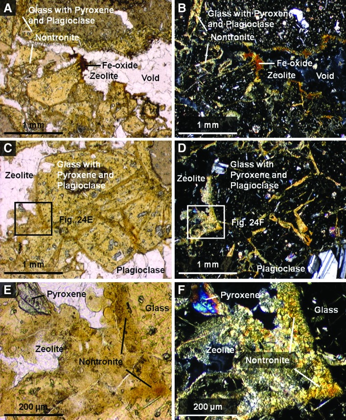
FIG. 24.
Plain light (left) and crossed-polarized light (right) images of magnified areas of thin section of sample 10 obtained with a Nikon Eclipse E600 polarizing microscope targeting main elements with matrix components labeled. These include matrix cements and clasts (A–D). (E and F) are close-ups of (C and D) to show the cross-cutting relationships of the cements to the clasts. The crossed-polarized images are slightly overexposed to bring out color variations between the first-order colors of zeolites, and clasts' glass matrix. Color images available online at www.liebertonline.com/ast

