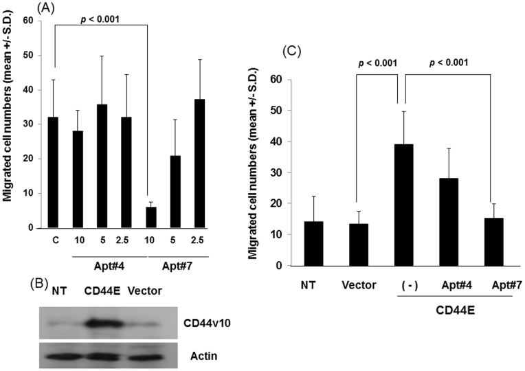
Figure 4. Inhibition of cell migration by aptamers against CD44 exon v10.
(A) HCC38 cells were harvested, washed and resuspended in RPMI1640-serum free media. Migration assays were performed using type I collagen (10 µg/ml) as an adhesive substrate in the lower compartment of Transwell by incubating at 37°C for 4 hours. The aptamers were added in both upper and lower chambers at the indicated concentrations in the text. Experiments were performed in triplicate. Statistical significance was calculated by Student’s two-tailed paired t-test. (B) MCF-7 cells were stably transfected with CD44E according to the methods described in Materials and Methods. MCF-7 cells transfected control vector, [MCF-7(pIRES2)], MCF-7 (CD44E), and parental MCF-7 cells (NT) were lysed and subjected to western blotting for the expression of CD44E using anti-CD44 exon v10 antibody followed by HRP-conjugated goat-anti-rabbit IgG. The signal was detected by ECL reactions. Actin was used as a loading control. (C) The migration of these cells was evaluated as described above in the presence or absence of aptamers against CD44 exon v10 at a concentration of 10 nM as described above. Experiments were performed in triplicate. Statistical significance was calculated by Student’s two-tailed paired t-test.

