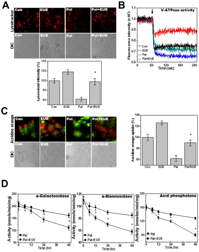
Figure 3. EUE regulates palmitate-reduced lysosomal activity.
Cells were treated with 500 µM palmitate in the presence or absence of 100 µg/mL EUE for 24 hours followed by exposure to 5 µM LysoTracker and image acquisition (A). Lysosomal fluorescence was quantified (A; lower). Lysosomal V-ATPase activity was measured as described in Materials and Methods (B). Acridine orange solution and valinomycin were added to cell monolayers and intravesicular H+ uptake was initiated by the addition of Mg-ATP (C); fluorescence was quantified at 24 hours (C; right). Cells were treated with 500 µM palmitate in the presence or absence of 100 µg/mL EUE for 0, 6, 12, 24, or 48 hours, and levels of α-galactosidase, α-mannosidase, and acid phosphatase were measured (D). * p<0.05, significantly different from palmitate-treated condition. DIC; differential interference contrast microscopy, Pal.; palmitate, EUE; Eucommia ulmoides Oliver extract.

