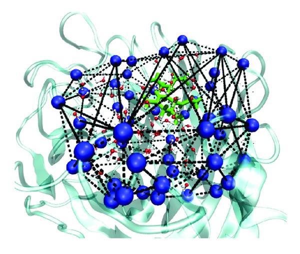
Figure 2.

The construction of convex hull for the binding site. Cα atoms of fifty residues which define the binding pocket are shown in blue ball. Not all sides of polyhedron are shown. Oseltamivir is colored in green. Water molecules are also presented.
