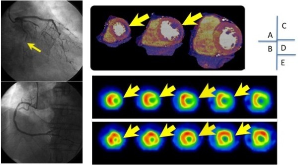
Figure 2.

Case (CAD = coronary artery disease, SPECT = single photon emission computed tomography). Case was a patient with asymptomatic CAD. Severe stenosis is seen in the left circumflex (panel A), and RCA is normal (panel B). Stress dual-energy imaging shows ischemia in the lateral wall (panel C), which correlated with the lateral wall ischemia seen on SPECT (panels D and E).
