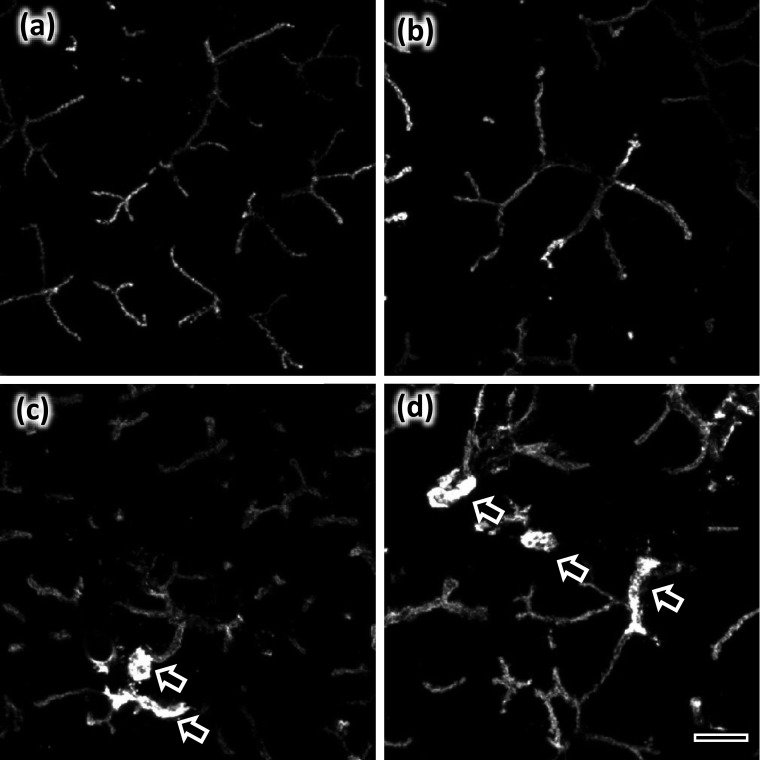
Fig. 4.
Subcellular distribution of AQP5 by laser confocal microscopy. Control (a) and IPR-administered (b) parotid glands. Control (c) and IPR-administered (d) submandibular glands. Projection images of 17 consecutive confocal images are shown. AQP5 is localized to the apical membrane including the intercellular canaliculi. No intracellular labeling was apparent. Arrows indicate intercalated ducts. Bar=10 µm.

