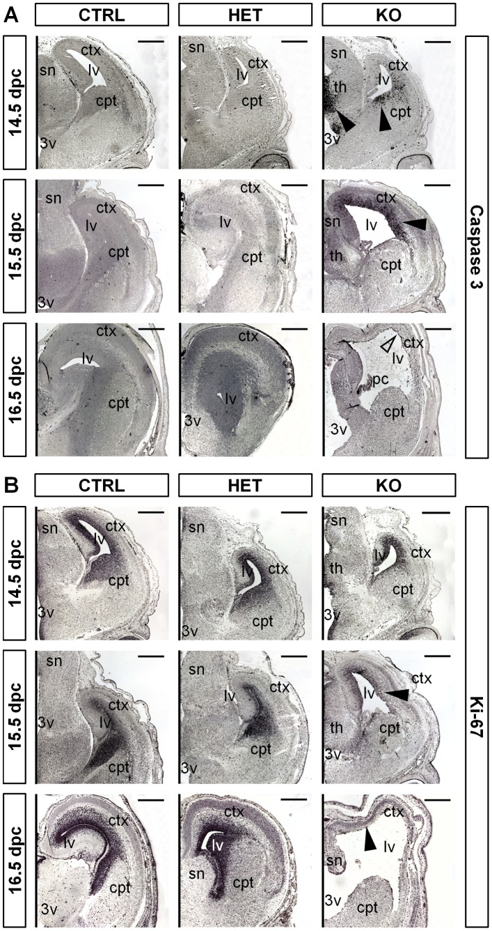
Figure 2. Brain malformations are initiated by massive apoptosis in the cortex.
(A) Immunostaining for Caspase-3 on paraffin-embedded coronal sections indicates prominent apoptosis in the proximal cortical layers and in the thalamic area of 14.5 dpc and 15.5 dpc KO embryos (black arrowheads). Remaining cortical tissue does not show apoptosis at 16.5 dpc and later stages (light arrowhead). (B) Immunostaining for Ki-67 shows initial decrease of proliferation at 14.5 dpc which is fully lost at 16.5 dpc in KO animals (black arrowheads). Control and HET animals retain strong Ki-67 signals in the proximal cortical layers at all indicated developmental stages. Scale bar equals 400 µm; ctx, cortex; th, thalamus; cpt, caudoputamen; sn, septal nuclei; 3v, third ventricle; lv, lateral ventricle.

