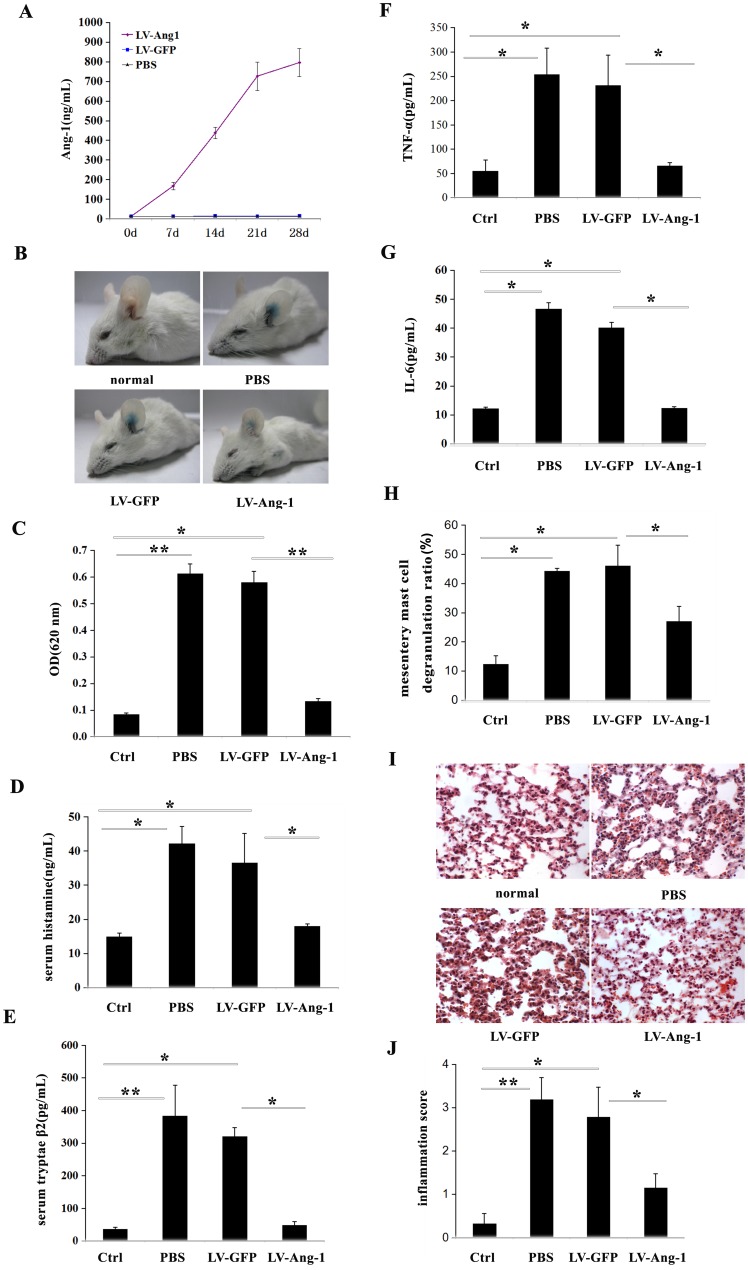
Figure 5. Ectopic expression of Ang-1 in vivo protected against IgE-dependent PCA and anaphylaxis shock.
A: The measurements of serum Ang-1 levels by ELISA in 7, 14, 21 and 28 days after intravenous injection of 1×10⁶ TU LV-Ang-1. Each data point indicates mean±SD of 5 mice. B and C: The anti-allergic effect was firstly assessed using IgE-DNP/DNP-BSA induced PCA mouse model. PCA mice showed ear Evans blue exudation (B), and the absorbance value was detected (C). D: Serum histamine of systemic anaphylaxis shock mice was determined through OPT-fluorometric assay as previously reported. Each data point indicates mean ±SD of 6 mice. E: ELISA kit detected serum tryptase-β2 of systemic anaphylaxis shock mices. Each data point indicates mean±SD of 6 mice. F and G: Peritoneal TNF-α and IL-6 levels in systemic anaphylaxis shock mices were detected by ELISA. Data indicate mean±SD from four independent experiments. H: In compound 48/80 induced systemic anaphylaxis shock model,mesentery mast cells degranulation was detected by toluidine blue staining. Quantification of mesentery mast cells degranulation by compound 48/80 was performed in a blinded fashion. I: Lung injury was detected by hematoxylin and eosin staining in systemic anaphylaxis shock mices. J: Statistical analysis of the lung injury score in systemic anaphylaxis shock mices by assessment infiltration of numerous polymorphonuclear leukocytes, interstitial spaces and hemorrhage, as described in materials and methods. It was performed in a blinded fashion. Each data point indicates mean±SD of 5 mice. *P<0.05.

