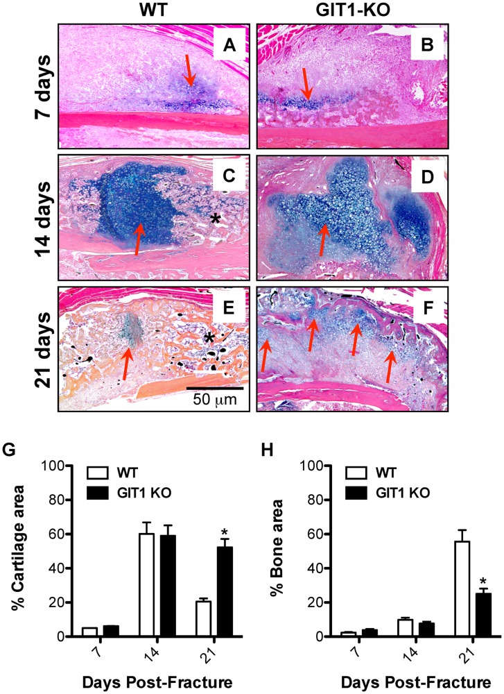
Figure 3. Cartilage persists and woven bone callus is delayed in GIT1 KO mice.
Histological analysis of fracture callus cartilage content was performed via Alcian Blue Hematoxylin/Orange G staining. Representative stains of calluses from WT and GIT1 KO mice at 1, 2 and 3 weeks post-fracture are displayed (A–F). Red arrows denote Alcian Blue-stained cartilagenous matrix and asterisks denote mineralized woven bone. Histomorphometry was performed on triplicate sections from multiple mice, with % Cartilage Area (G) and % Bone Area (H) quantified. Bars represent % Area (cartilage or bone) +/− SEM (*p<0.05, N = 3).

