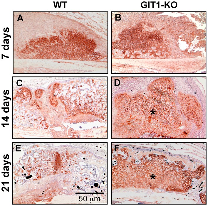
Figure 4. Type 2 collagen-containing matrix persists in GIT1 KO mice.
Tissue sections cut from WT and GIT1 KO mice were analyzed for COL2A1 content using an immunohistochemistry approach. Representative stains at 7, 14 and 21 days post-fracture are depicted, with asterisks denoting areas within the callus at 2 and 3 weeks post-fracture in GIT1 KO mice (D and F respectively) that have more robust/persistent staining.

