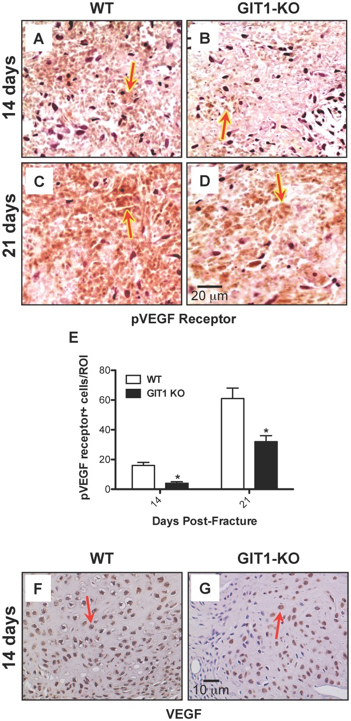
Figure 9. VEGF signaling is reduced in GIT1 KO mice.
Phospho-VEGF receptor immunostaining was performed on fracture calluses from WT and GIT1 KO mice at 2 and 3 weeks post-fracture. Panels A-D depict representative staining profiles, with Phospho-VEGF receptor-positive cells staining red as indicated by red arrows. Histomorphometry was performed to quantify the number of Phospho-VEGF receptor-positive cells per unit callus area (E). Data is presented as the mean number of positive cells per unit area (i.e. region of interest) +/− SEM (*p<0.01, N = 3). Additionally, immunohistochemistry was performed of assess VEGF levels in fracture calluses from WT (F) and GIT1 KO mice (G). Representative histological sections of calluses at 2 weeks post-fracture are presented, depicting reduced expression in KO mice. VEGF positive cells are stained reddish-brown as indicated by red arrows.

