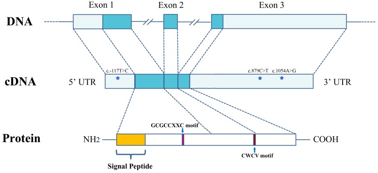
Figure 1. The structure of the PyIGFBP gene and protein.
The gene contains three exons. The 5′ and 3′ UTR (light blue) and exons (blue) are shown relative to their lengths. The location of the three SNPs (c.-117T>C, c.879C>T and c.1054A>G) is indicated with a star. The position of the GCGCCXXC and CWCV motifs is indicated with an arrow, respectively.

