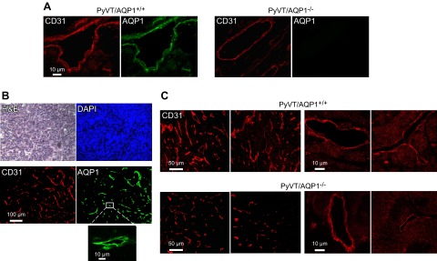
Figure 2.

Tumor microvasculature in primary breast tumors of female AQP1+/+ and AQP1−/− PyVT mice at 98 d. A) CD31 (red) and AQP1 (green) immunofluorescence showing colocalization in tumor of AQP1+/+ PyVT mouse. B) H&E, DAPI, CD31 and AQP1 in tumor of AQP1+/+ PyVT mouse showing absence of AQP1 expression in tumor cells. C) Examples of CD31 immunofluorescence of breast tumors from 2 AQP1+/+ and AQP1−/− PyVT mice at low (left panels) and high (right panels) magnifications.
