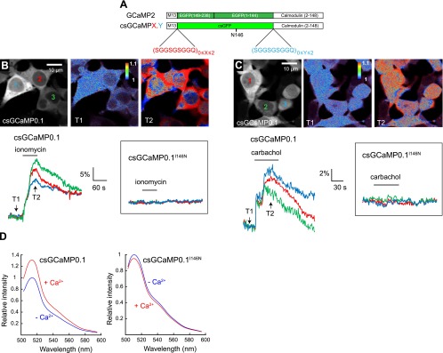
Figure 5.

csGFP reports activation of a calcium-sensing domain. A) Domain structure of constructs derived from the GCaMP2 calcium sensor and incorporating a CFP-derived csGFP, as well as variable numbers of a nonapeptide spacer. B, C) Fluorescence images show the distribution of the csGCaMP0.1 sensor in the cytoplasm and nucleus of HEK cells (grayscale), and fluorescence increase of this sensor (pseudocolor) elicited by application of ionomycin (4 μM; B) or carbachol (300 μM; C). Traces illustrate csGCaMP0.1 responses measured in indicated cells (colored numbers in top left panels) and the absence of detectable response of the csGCaMP0.1 I146N mutant on application of these drugs (insets). D) Emission spectra of csGCaMP0.1 and csGCaMP0.1 I146N in the presence or absence of calcium (1 mM), measured in a cell-free assay following bacterial expression of the constructs.
