
Figure 7.
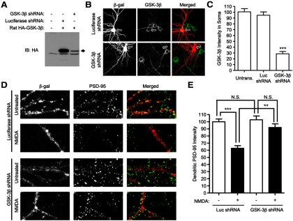
RNAi knockdown of GSK-3β reduces NMDA-induced loss of synaptic PSD-95. A, COS-7 cells were cotransfected with indicated expression vectors, and immunoblotted 60 h later for HA-GSK-3β. B, Cultured hippocampal neurons at DIV 15 were cotransfected with luciferase- or GSK-3β-shRNA constructs plus β-gal marker (4:1 shRNA: marker). Three days later, neurons were fixed and double-stained with mouse β-gal and rabbit GSK-3β antibody. Arrows point to soma of transfected neurons. C, Bar graph showing soma staining intensity of GSK-3β, normalized to untransfected cells (n = 12, 12, and 13 from left to right; t test, ***p < 0.001 compared with untransfected neurons or neurons transfected with luciferase shRNA). D, Cultured hippocampal neurons at DIV 15–16 were cotransfected with luciferase-shRNA, or GSK-3β-shRNA, plus marker β-gal. Three days later, transfected neurons were either untreated or treated with 75 μm NMDA for 10 min, and then double-labeled for β-gal and PSD-95. E, Bar graph showing dendritic immunostaining intensity of PSD-95, normalized to untransfected cells. Statistical analysis was performed by one-way ANOVA with a Bonferroni post hoc test for the indicated comparisons (n = 11 neurons for each condition; **p < 0.01, ***p < 0.001).