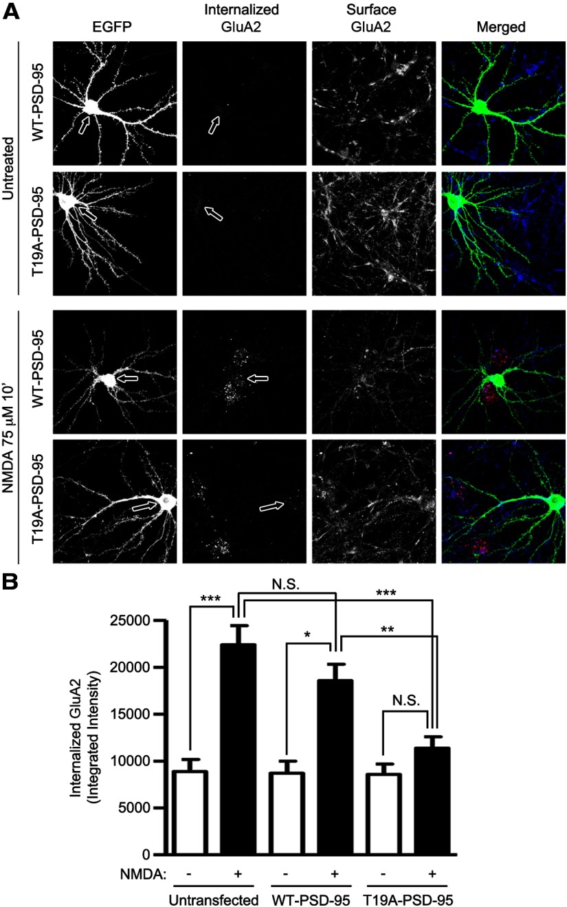
Figure 9.
Overexpression of PSD-95-T19A inhibits NMDA-induced GluA2 internalization. A, Cultured hippocampal neurons were transfected with EGFP, WT-PSD-95-EGFP, or T19A-PSD-95-EGFP at DIV 16. Three days later (DIV 19), surface GluA2 was live-labeled with GluA2-N antibody, washed, and then left untreated or treated with 75 μm NMDA for 10 min at 37°C. Internalized GluA2 receptors and remaining surface GluA2 receptors were visualized with Alexa-568 (red), and Alexa-647 (blue) secondary antibody, respectively (see Materials and Methods). Transfected neurons were identified with GFP-channel (green). Arrows point to the cell bodies of transfected neurons. B, Bar graph shows mean ± SEM. of integrated intensity of internalized GluA2 receptor. Statistical analysis was performed by one-way ANOVA with a Bonferroni post hoc test for the indicated comparisons (n = 14, 43, 9, 20, 8, and 20 from left to right; *p < 0.05, **p < 0.01, ***p < 0.001).

