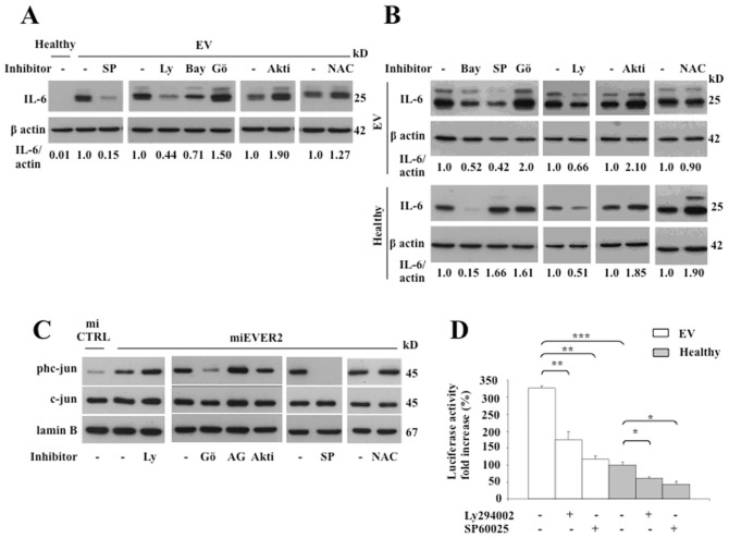
Figure 5. EVER2 controls NF-κB and JNK/AP-1 pathways.
(A–D) Cell lines were left untreated (-) or were treated with specific inhibitors of JNK (SP60025 (SP; 20 µM)), PI3K (Ly294002 (Ly; 15 µM)), IKKβ (Bay11-7082 (Bay; 5 µM)), PKCα (Gö6976 (Gö; 0.5 µM)), AKT1/2 (Akti; 2 µM), EGF receptor (AG1478 (AG; 12.5 µM) or N-acetylcysteine (NAC; 10 µM) as indicated. EV and Healthy cell lines were left unstimulated (A) or were stimulated with TNF (B) in the presence of brefeldin A for 16 hours. Whole-cell lysates were assayed for intracellular IL-6 by western blotting. The data shown are from one experiment representative of two independent experiments carried out. The IL-6 and actin bands on western blots were quantified by densitometry. Results are reported as the ratio of IL-6 to actin. In panel A, the ratio obtained for untreated EV cells was set to 1. In panel B, the ratio was set to 1 for each cell line in the absence of inhibitors. (C) miEVER2 and miCTRL cell lines were left untreated (-) or were treated with inhibitors specific as indicated. We analyzed the levels of c-jun or its phosphorylated form (phc-jun) in nuclear extracts by western blotting. The results shown are representative of two independent experiments. (D) EV and Healthy cell lines were transiently transfected with 0.3 µg/well HPV5-flLCR/luc or HPV5-minLCR/luc. Cells were assayed for luciferase activity. Results are reported as fold-increases over the level obtained with HPV5-minLCR/luc. Values obtained with the unstimulated Healthy cell line are set to 100%. The data shown are means ± SD of three independent experiments. *, P<0.05; **, P<0.01; ***, P<0.001.

