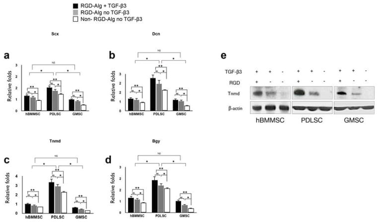
Figure 3. Specific gene expression and underlying molecular pathway for tendon regeneration in vitro.
Expression level (in fold changes) of (a) Scx, (b) Dcn, (c) Tnmd, and (d) Bgy genes for each encapsulated stem cell population after 4 weeks of culturing in induction media in vitro evaluated by RT-PCR. Data were normalized by the Ct of the housekeeping gene GAPDH and expressed relative to the expression level for the same gene at day 1. (e) Western blot analysis showing changes in the levels of expression of regulators of tenogenesis of MSCs. The level of Tnmd is elevated in the encapsulated MSCs in RGD-containing alginate microspheres in the presence of TGF-β3. MSCs encapsulated in non-RGD-coupled alginate microspheres in the presence of TGF-β3 exhibited very modest levels of Tnmd expression, while specimens in the absence of TGF-β3, encapsulated in non-RGD-coupled alginate microspheres failed to express Tnmd. *P <0.05, NS= not significant.

