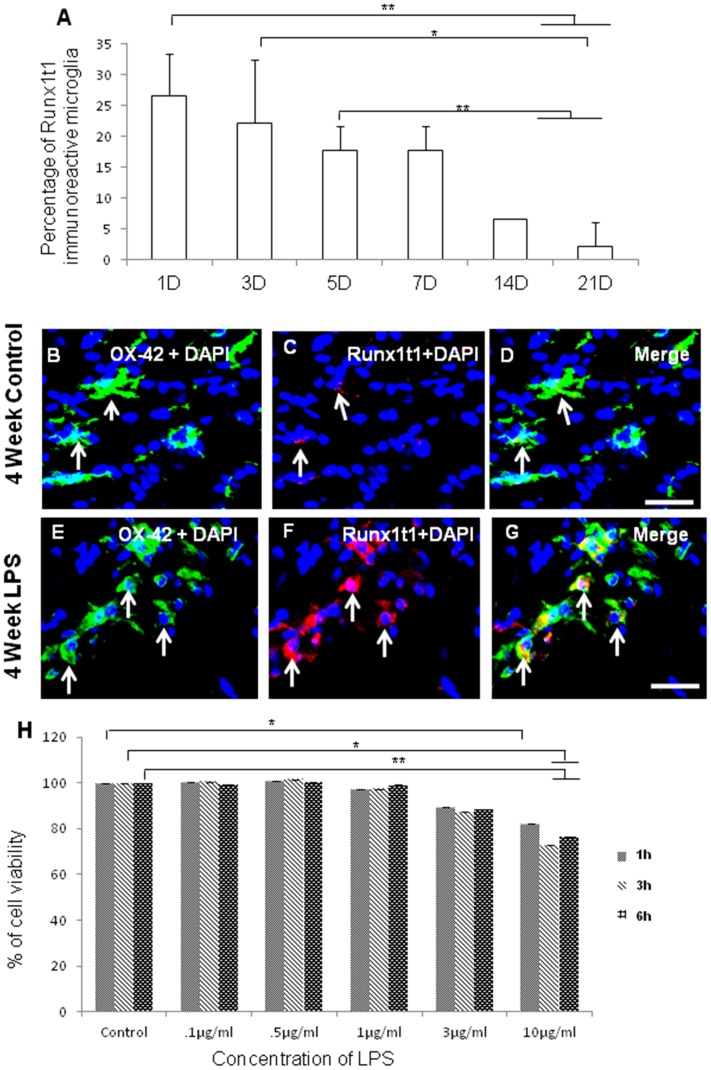
Figure 2. Quantitative analysis showing the percentage of Runx1t1 immunoreactive microglia in rat brain.
A. Quantitative analysis shows that number of Runx1t1 immunoreactive microglia decreases with age in normal rats. Significant differences between groups (1D vs14D, 1D vs 21D, 3D vs 21D, 5D vs 14D, 5D vs 21D) are indicated by *p<0.05, **p<0.01 (n = 3). B–G. Confocal images showing the colocalization of Runx1t1 (C,F; red) and OX42 (B,E; Green) in control (B–D) and LPS-treated (E–G) activated microglia from the corpus callosum of 4 week rat brain. Nuclear translocation of Runx1t1 is evident in LPS-treated activated microglia (F;arrows). Nuclei are stained with DAPI. Scale bars: 20 µm. H. MTS assay shows the effect of various concentrations of LPS (0.1 µg/ml–10 µg/ml) on the viability of BV2 microglial cells. Treatment of these cells with different concentrations of LPS (0.1 µg/ml–10 µg/ml) for 1 h, 3 h and 6 h revealed that LPS did not affect the cell viability up to 1 µg/ml concentration. Cell viability was decreased with further increase in the concentration. Significant differences between groups (Control vs LPS (3 µg/ml), Control vs LPS (10 µg/ml) are indicated by *p<0.05, **p<0.01 (n = 3).

