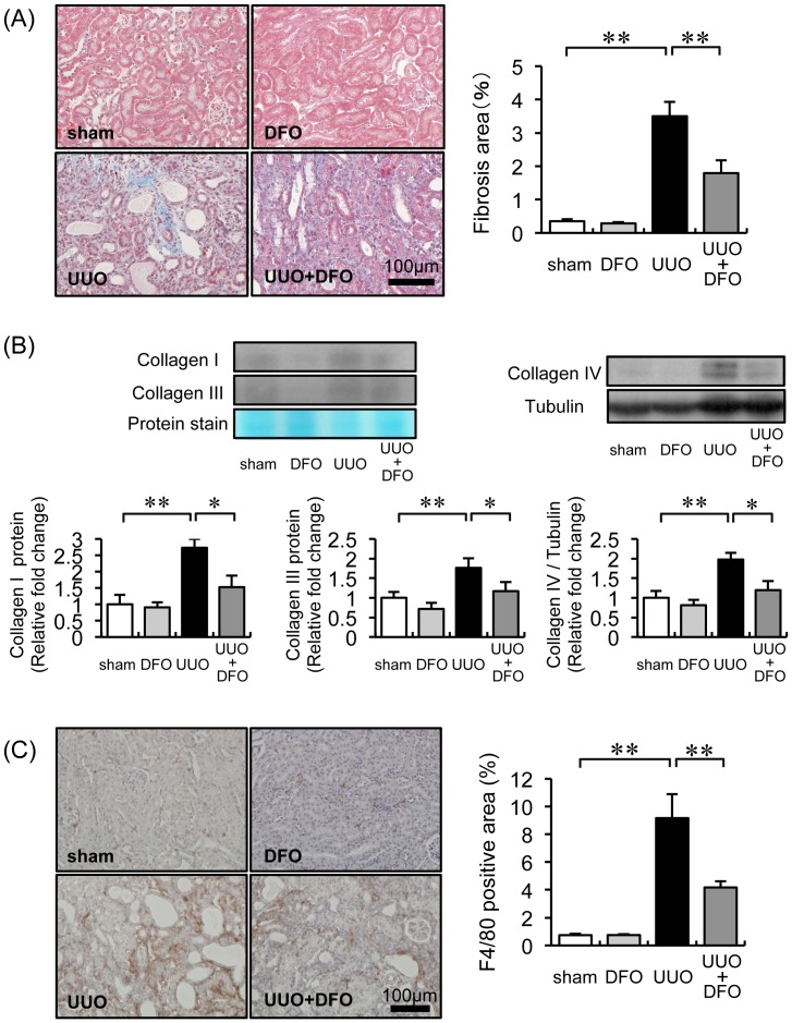
Figure 1. Effect of DFO on UUO-induced renal interstitial fibrosis, collagen expression, and macrophage infiltration.
(A) Representative histological findings in tissue stained with Masson’s trichrome 7 days after surgery. (B) Quantitative analysis of renal interstitial fibrosis at day 7. Sham+VEH (white bar), sham+DFO (light gray bar), UUO+VEH (black bar), and UUO+DFO (gray bar). Results are expressed as the mean ± SEM. *P<0.05, **P<0.01. n = 12 per group. (C) Analysis of the renal expression of collagen IA, collagen IIIA, and collagen IV. Upper panels: representative immunoblotting for collagen IA, collagen IIIA, and collagen IV. Lower panels: quantitative analysis of collagen IA, collagen IIIA, and collagen IV expression normalized to protein staining or tubulin. Results are expressed as the mean ± SEM. *P<0.05, **P<0.01. n = 12 per group. (D) Representative immunohistochemistry with F4/80 antibody 7 days after surgery. (E) Quantitative analysis of the F4/80-positive area within the interstitial area. Results are expressed as the mean ± SEM. *P<0.05, **P<0.01. n = 12 in each group.

