
Figure 5.
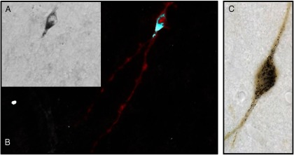
Double labeling of mRNA and protein in the same cell. A, GnRH. The image on the left shows GnRH mRNA using a biotinylated riboprobe stained with immunoperoxidase detection of the biotin in black. B, Inversion converts the black product to white, and staining using immunofluorescence in red shows that the same cell that possessed mRNA also possesses the product of that mRNA, GnRH peptide. C, Tyrosine hydroxylase. As in panels A and B, but the cell in this image has its mRNA stained black and the protein tyrosine hydroxylase in brown.