Figure 7.
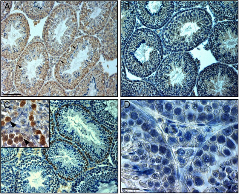
Immunohistochemical labeling of mouse testis tissue with a mouse monoclonal antibody against the protein p62 (brown staining), a marker for autophagy. Examples of labeled spermatogonia and primary spermatocytes are indicated by arrowheads. A, Conventional immunohistochemical staining procedure, using normal goat serum and a biotinylated goat-antimouse secondary antibody. Note that except for the cytoplasm of the elongating spermatids all cells seem to stain. B, Omission of the primary p62 antibody, otherwise using the same procedure as in panel A; the staining in spermatogonia and spermatocytes has more or less disappeared, although background staining in most cells remains present. C, Normal goat serum has been replaced by a preincubation with goat-antimouse Fab fragments, and the biotinylated secondary goat-antimouse antibody has been replaced by a F(ab)2-labeled biotinylated goat-antimouse antibody; now there is specific brown staining in the spermatogonia and primary spermatocytes only and an absence of any background staining. The inset in panel C shows a detail of this. D, As negative control, the p62 antibody has been replaced by IgG of the same class as the primary antibody; otherwise the procedure is exactly as in panel C. Scale bars: panel A, 42 μm; panel D, 10.5 μm.

