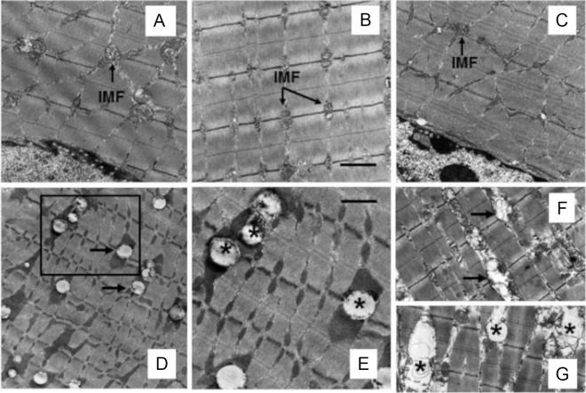
Figure 2.
A and B, Representative transmission electron micrographs of gastrocnemius muscle from mice fed an NCD without (A) or with (B) nicotine exhibit normal myofibrillar architecture and sarcomeric pattern with abundant normal-appearing IMF mitochondria and no IMCL accumulation. C, Appearance of muscle fiber in mice fed an HFD is similar to that seen in mice on an NCD in the absence (A) or presence (B) of nicotine. D, Representative electron micrograph shows IMCL accumulation in close association with IMF mitochondria (arrow). E, A higher-magnification view of the area marked in D showing IMCL accumulation (asterisk). F and G, Nicotine plus HFD also causes mitochondrial vacuolization (F; arrow) and mitochondrial swelling with broken cristae (G; asterisk). Scale bar, 1 μm (A–D, F, and G) and 0.4 μm (E). Data are representative of 4 mice in each group.

