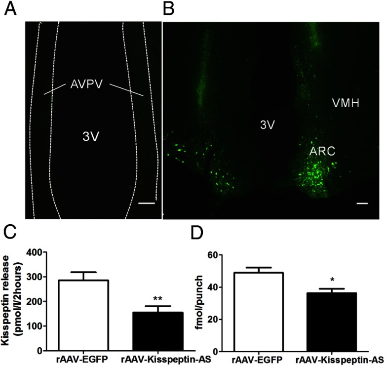
Figure 1.
Validation of transgene expression and vitro kisspeptin knockdown. Representative images showing rAAV-EGFP spread in the (A) AVPV and (B) ARC (B). VMH, ventromedial hypothalamus; 3V, third ventricle. Scale bars represent 100 μm. C, Kisspeptin release from the RIN 1056a cell line stably transfected with Kiss1 after transient transfection with EGFP (control) or Kisspeptin-AS after 2 hours of incubation. Data are presented as mean ± SEM (n = 8); **, P < .01 vs kisspeptin-AS. D, The effect of intra-Arc rAAV-EGFP and rAAV-Kisspeptin-AS administration on kisspeptin-IR in the ARC. Quantification of kisspeptin-IR in the ARC of female rats by RIA (n = 12–14). Data are shown as mean ± SEM; *, P < .05.

