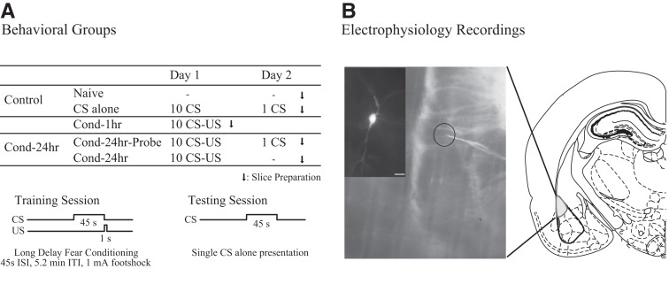
Figure 1.
Experimental design used to study learning-related changes in the amygdala. (A) Behavioral groups. Rats were divided into five groups: two control groups (naive [N = 12]) and CS-alone [N = 4]) and three experimental groups (Cond-1hr [N = 8], Cond-24hr-Probe [N = 9], and Cond-24hr [N = 3]; see Materials and Methods for details). (B) Electrophysiological recordings. Right panel is a schematic of a typical coronal brain slice showing the location of the lateral amygdala (LA). Left panel is a photomicrograph of a brain slice showing the location of a typical recording electrode (inset is a neurobiotin-filled LA pyramidal neuron; scale, 40 µm).

