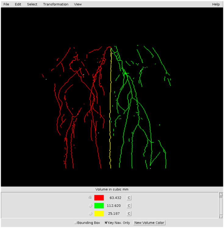
Fig. (2). Screenshot of MRA image in VolCal.

The screenshot shows a skeletonized image in VolCal and how the user can choose blood vessel trees and highlight them in different colors. The user has also the option of picking single blood vessels. In the lower part of the window the total volumes for the vessels of a given color are shown, in this case the ischemic leg is shown in red, the non-ischemic leg in green and the tail in yellow.
