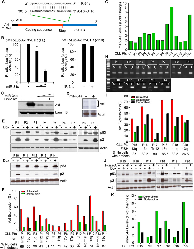
Figure 1. miR-34a targets Axl expression in primary CLL B-cells.
A. Alignment of Axl 3’-UTR with miR-34a sequence. B. miR-34a targets Axl 3’-UTR. HEK293 cells were co-transfected with a luciferase reporter plasmid DNAs containing the entire Axl 3’-UTR with a putative miR-34a-binding site (full-length) or without the miR-34a-binding site [Axl 3’-UTR (-110)] and increasing amounts of miR-34a mimic or sc- miR as control. After 24 hour, luciferase activity was measured in the cell lysates. Experiments were repeated twice in triplicates. Data were normalized and presented as mean values with standard deviations. C. Exogenous miR-34a reduces Axl expression. HEK293 cells were co-transfected with an Axl expressing plasmid DNA containing the putative miR-34a binding site in the 3’-UTR and increasing amounts of miR-34a or sc-miR for 24 hours. Cells were examined for the expression of Axl by Western blot using a specific antibody to Axl. Lamin B was used as a loading control. D. Introduction of miR-34a mimic reduces Axl expression in primary CLL B-cells. Purified CLL B-cells were transfected with miR-34a mimic or sc-miR as control using B-cell specific nucleofection reagent (Amaxa). After 24 hours, cell lysates were analyzed for the expression of Axl by Western blots using a specific antibody. Actin was used as a loading control. Representative result of CLL B-cells from 3 different CLL patients is shown. E. Doxorubicin activates p53. Lysates from purified CLL B-cells treated with doxorubicin or left untreated for 16 hours were analyzed for the expression of p53 and its downstream target p21 in Western blots using specific antibodies. Actin was used as a loading control. CLL B-cells from various CLL patients are indicated by arbitrary numbers (P1-P14). F. Impact of doxorubicin on Axl expression. CLL B-cells from the same CLL patients’ cohort (P1-P14) studied above (panel E) were treated with doxorubicin or left untreated for 16 hours. Axl expression was determined by flow cytometry using a specific antibody. Percent of cells with FISH detectable chromosomal abnormalities are indicated. G. Doxorubicin treatment activates miR-34a expression in CLL B-cells. Total RNA was extracted from untreated and doxorubicin-treated CLL B-cells and miR-34a levels were measured by qRT-PCR using primers specific for mature miR-34a and normalized using the 2-∆∆Ct method relative to U6-snRNA (RNU6B). Relative miR-34a expression levels in untreated vs. doxorubicin-treated CLL B-cells are presented as “fold changes” determined as mean of triplicate values. CLL B-cells studied here were chosen from the same cohort of CLL patients depicted and studied above in panels E&F. H. Methylation specific-PCR analysis of the miR-34a promoter in CLL B-cells. DNA isolated from purified CLL B-cells of the CLL patients (P1 - P3, P5, P7 - P14) whose miR-34a levels were studied above (panel G) were subjected to bisulfite conversion and analyzed by methylation specific-PCR (MSP) with primers specific for methylated (M) and unmethylated (U) miR-34a promoter DNA. Amplified PCR products were run on a 1% agarose gel along with a standard DNA ladder (Invitrogen; 1st lane from left) to ascertain the size of the PCR products. I. In vitro Fludarabine treatment reduces Axl expression on CLL B-cells. CLL B-cells from CLL patients (P15 – P20) were treated with fludarabine (F-ara-A) for 16 hour or left untreated and Axl expression was determined by flow cytometry as described above. Doxorubicin was included as a positive control. J. Fludarabine activates p53. Cell lysates prepared from untreated, doxorubicin-treated and F-ara-A-treated CLL B-cells from CLL patients (P15 – P20) were examined for the expression of p53 and p21 in Western blot analysis. Actin was used as a loading control. K. Fludarabine upregulates miR-34a expression. Total RNA was extracted from untreated, doxorubicin-treated and F-ara-A-treated CLL B-cells from the same CLL patients used above (P15 – P20). Mature miR-34a levels were measured by qRT-PCR and presented as “fold changes” as described in panel G.

