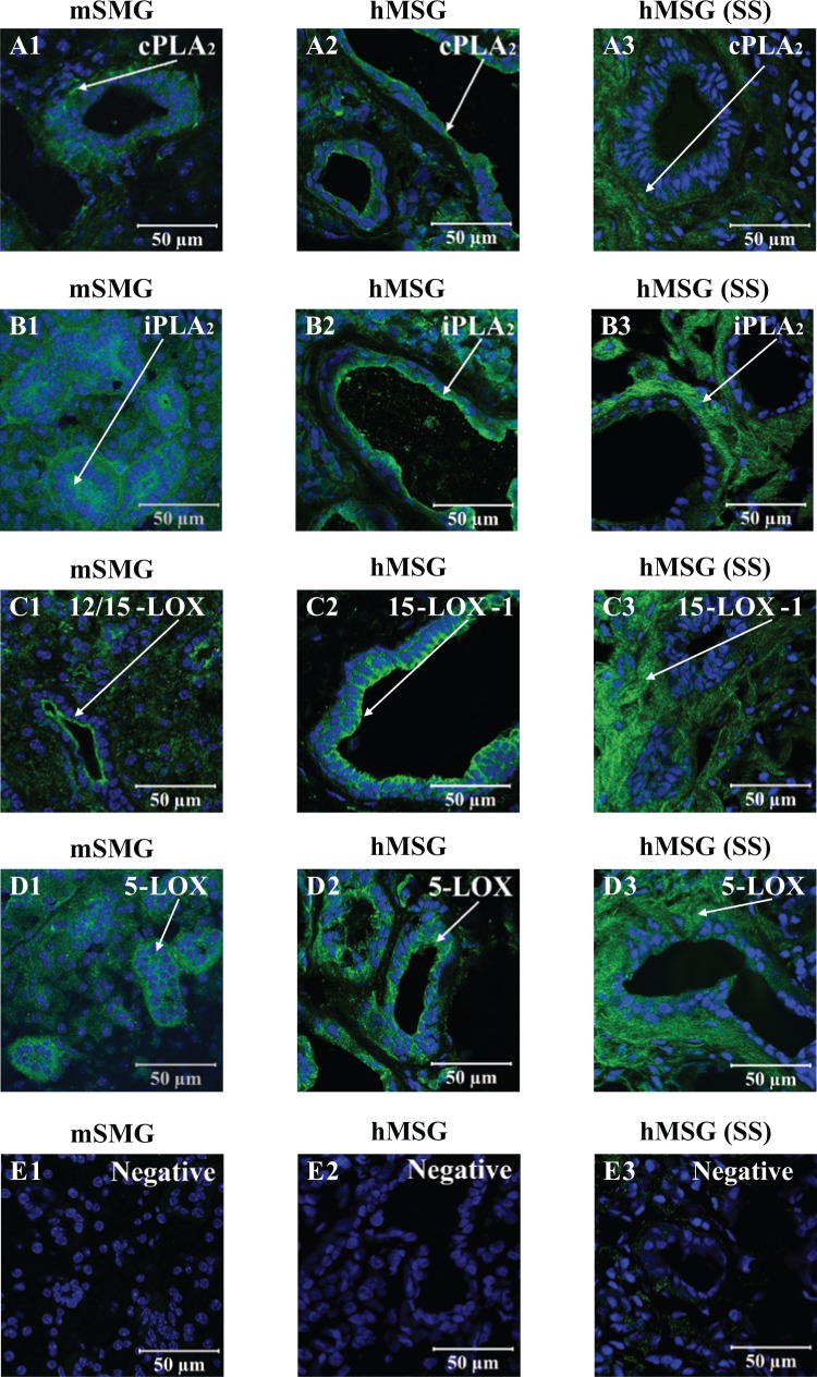
Figure 3.
Resolvin D1 (RvD1) biosynthetic machinery localization in salivary glands with and without Sjögren’s syndrome (SS). Frozen sections from mouse submandibular gland and human minor salivary glands with and without SS were subjected to immunofluorescent staining as described in the Appendix Materials & Methods. Sections were stained (A1, A2, and A3; green) with goat anti-rabbit anti-cPLA2, (B1, B2, and B3; green) goat anti-rabbit anti-iPLA2, (C1, C2, and C3; green) goat anti-rabbit anti-12/15-LOX, (D1; green) goat anti-sheep anti-15-LOX type-1, and (D2 and D3; green) goat anti-rabbit anti-5-LOX, followed by Hoechst or Propidium Iodide Nucleic Acid Stain (all images; blue). Images were obtained and analyzed with a Zeiss LSM 510 confocal microscope. White arrows indicate enzyme localization.

