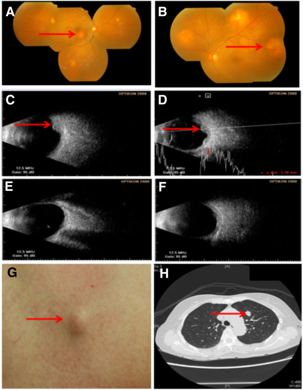
Figure 1.
Choroid and skin metastases of primary clear cell adenocarcinoma of lung. (A-F) Ophthalmology images; (A,C,E) right eye, (B,D,F) left eye. (A,B) Fundus appearance before treatment (arrows point to lesions); (C,D) ultrasound scan of the same eyes as in (A,B) before treatment (arrows point to lesions); (E,F) lesion resolution by ultrasound scan of the same eyes as in (A,B) after treatment. (G) Appearance of skin metastasis (arrow). (H) Chest computed tomography scan showing left upper lobe mass (1.3cm in diameter) (arrow).

