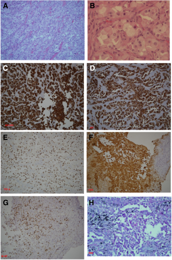
Figure 2.
Histopathological findings of primary clear cell adenocarcinoma of the lung. Hematoxylin and eosin staining showed that the main tumor was infiltrated by pleomorphic clear tumor cells with foamy cytoplasm and distinct nucleoli ((A), 100×; (B) 400×). Positive immunohistochemical staining results included pancytokeratin AE1/AE3 (C), cytokeratin 7 (D), thyroid transcription factor 1 (E), Ki-67 (G), carcinoembryonic antigen (F) and periodic acid Schiff (H). Negative immunohistochemical stainings included HMB-45, Hep-par-1, transcription factor E3, α-inhibin, S-100 and CDX2 (data not shown).

