
Figure 3.
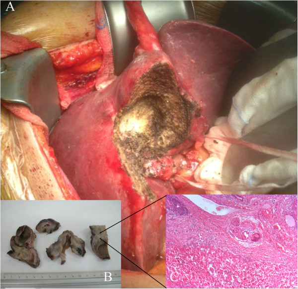
Specimen examination. A) A typical partial resection of the 5th Segment en-bloc with the gallbladder. Intrahepatic position of the gallbladder. B) Macroscopic details of the pathologic specimen: intrahepatic position of the gallbladder. C) Microscopic detail of the specimen (Zoom 10 X): chronic inflammatory cells (giant plurinucleated cells) mixed with hepatic regeneration nodes in the context of a granuloma under the gallbladder bed, effect of intrahepatic perforation. (Courtesy of Dr. Loredana Villari. Pathology Institute. Vittorio-Emanuele Hospital. Catania.).