
Fig. 2.
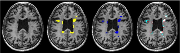
Axial view of 3DT1 image and different lesion masks for a 47-years old, male RRMS patient scanned at 3 T. From left to right: Image with no lesion masks. Image with precise manual lesion masks (yellow). Image with nearest neighbor registered lesion masks (dark blue). Image with trilinearly registered lesion masks with different thresholds: light blue: threshold = 0.2; red: threshold = 0.5; pink: threshold = 0.8.