
Fig. 5.
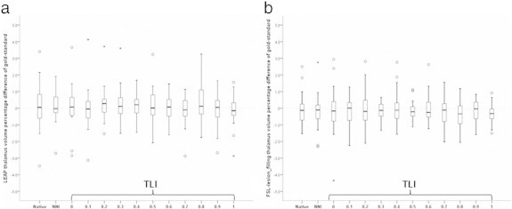
Boxplots of the relative error in mean bilateral thalamic volumes from segmentation of images after filling lesions obtained with different lesion masks using LEAP for lesion filling (a) and FSL lesion_filling (b). The thalamic volumes are reported as percentual difference relative to the thalamic volume obtained after filling the gold-standard (manual) lesion masks and then applying FSL-FAST segmentation. Native = native (unfilled) images. NNI = after filling with nearest-neighbor co-registered masks. TLI = after filling with trilinearly co-registered masks and then thresholding with the indicated value. These graphs are similar to the behavior of the other DGM structures both with LEAP lesion filling as well as with FSL-lesion_filling.