
Fig. 7.
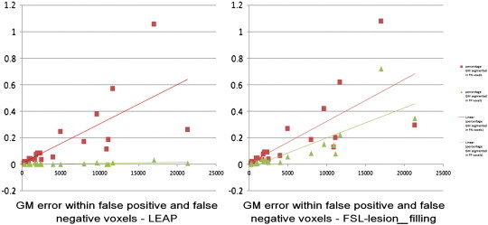
Percentage of grey matter (GM) segmented within the false negative and false positive voxels of the images filled with nearest-neighbor registered masks (NNI) after lesion-filling with LEAP (left) and FSL-lesion-filling (right) as a function of the gold-standard lesion volume. In red with square markers is depicted the percentage of GM segmented within the false negative voxels and in green with triangular markers the percentage of GM segmented within the false positive voxels.