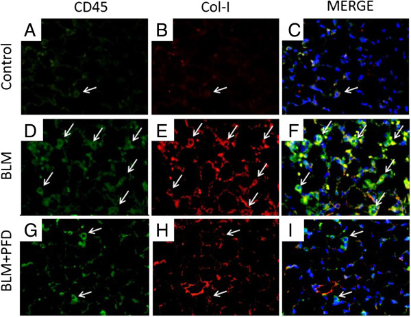
Figure 4.
Immunohistochemical analyses. Immunohistochemical analyses for CD45 (green; A, D, and G) and collagen-I (Col-I; red; B, E, and H) staining were performed with lung sections on day 14 of BLM treatment with or without pirfenidone. Merge (yellow; C, F, and I) is co-staining for CD45 and Col-I, indicating bone marrow-derived fibrocytes. Fibrocytes are indicated with arrows. The increased numbers of fibrocytes expressing CD45 and Col-I observed in BLM-treated mice were attenuated by pirfenidone treatment. Nuclei were counterstained with 4′,6-diamidino-2-phenylindole. Original magnification, ×40. PFD, pirfenidone.

