. 2014 Feb 25;3:e01612. doi: 10.7554/eLife.01612

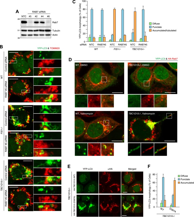
Figure 8. Rab7 is involved in autophagosome fusion during mitophagy.

(A) Total cell lysates from HCT116 treated with control (NTC) or RAB7A siRNA were analyzed by immunoblotting. (B) The indicated cells stably expressing YFP-LC3 (green) and mCherry-Parkin were treated with control (NTC) or Rab7_#5 siRNA. After 3 hr valinomycin treatment, cells were analyzed by immunofluorescence microscopy with anti-TOMM20 antibody (red). Z-stacks of confocal images are shown. Magnified images are also shown. Scale bars, 10 μm. (C) YFP-LC3 morphologies of cells in (B) were quantified. Percentages of cells harboring diffuse, punctuate or accumulated/tubulated YFP-LC3 are shown. Data and error bars were obtained from at least 50 cells in each of three independent replicates. (D) The indicated cells stably expressing YFP-LC3 (green), mCherry-Parkin, and 2HA-Rab7 (Red) were treated with or without valinomycin for 3 hr and analyzed by immunofluorescence microscopy with anti-HA antibody. Magnified images are also shown. Scale bars, 10 μm. (E) YFP-LC3 and mCherry-Parkin stably expressing TBC1D15−/− cells in the presence of HA-tagged TBC1D15 WT or the D397A mutant were treated with valinomycin for 3 hr. Cells were subjected to immunofluorescence microscopy with anti-HA antibody. Scale bars, 10 μm. (F) The YFP-LC3 morphology of cells in (E) was quantified. The error bars represent ±SD from three independent replicates. Over 50 cells were counted in each replicate.

DOI: http://dx.doi.org/10.7554/eLife.01612.024

Figure 8.

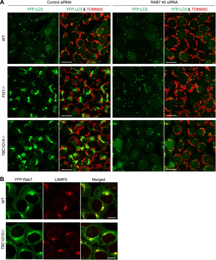
Figure 8—figure supplement 1. Rab7 is involved in LC3 accumulation of FIS1−/− and TBC1D15−/− cells.

Figure 8—figure supplement 1.

(A) Low magnification images of cells stably expressing YFP-LC3 and mCherry-Parkin treated with valinomycin for 3 hr. Cells were subjected to immunofluorescence microscopy with anti-TOMM20 antibody. Z-stacks of confocal images are shown. Scale bars, 20 μm. (B) The indicated cells stably expressing YFP-Rab7 and mCherry-Parkin were analyzed by immunostaining with anti-GFP and anti-LAMP2 antibodies. Scale bars, 10 μm.
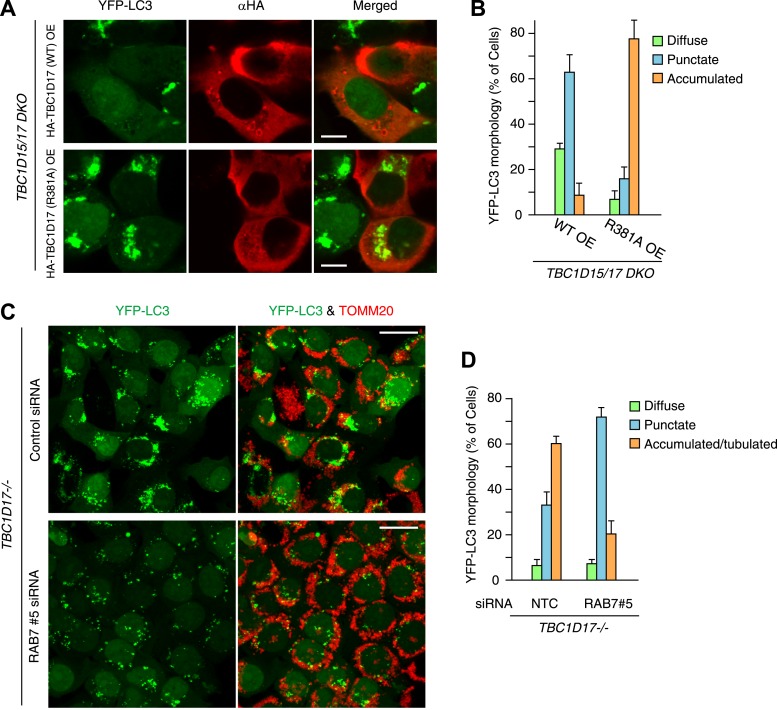
Figure 8—figure supplement 2. TBC1D17 GAP activity is important for LC3 accumulation through Rab7.

Figure 8—figure supplement 2.

(A) HA-tagged TBC1D17 WT or the R381A mutant was transiently overexpressed (OE) in TBC1D15/17 DKO cells stably expressing YFP-LC3 and mCherry-Parkin. The cells treated with valinomycin for 3 hr were subjected to immunofluorescence microscopy with anti-HA antibody. Scale bars, 10 μm. (B) The YFP-LC3 morphology of cells in (A) was quantified. The error bars represent ±SD from three independent replicates. Over 50 cells were counted in each replicate. (C) TBC1D17−/− cells stably expressing YFP-LC3 (green) and mCherry-Parkin were treated with control (NTC) and Rab7_#5 siRNAs for 48 hr. The cells treated with valinomycin for 3 hr were then subjected to immunofluorescence microscopy with anti-TOMM20 antibody (red). Z-stacks of confocal images are shown. Scale bars, 20 μm. (D) The YFP-LC3 morphology of cells in (C) was quantified. The percentages of cells harboring diffuse, punctate or accumulated/tubulated YFP-LC3 were quantified. The error bars represent ±SD from three independent replicates. Over 50 cells were counted in each of three replicate.