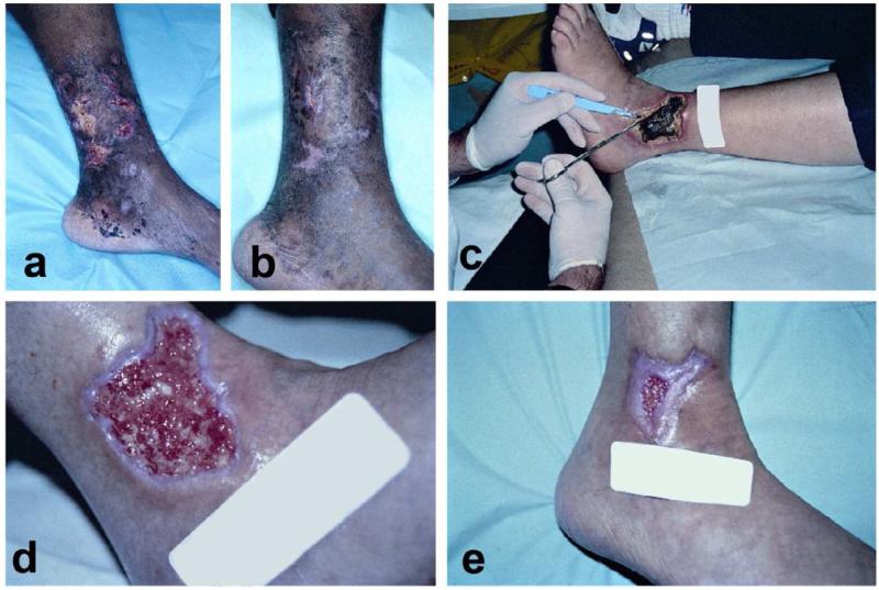
Figure 3. Examples of vasculitic ulcers.
A-B: Biopsy proven periarteritis nodosa with destruction of medium-sized vessels. Notice the slight microlivedo pattern surrounding the ulcer as well as the necrotic appearance of some of the ulcers. Treatment included methotrexate, sulfamethoxazole for pnuemocystis pneumonia (PCP) prophylaxis, and with a gel that promoted a moist wound environment and helped with autolytic debridement. C-E: This patient has an established diagnosis of Wegners granulomatosis c-ANCA positive small-medium sized vessel vasculitis. C: This large necrotic ulcer needed surgical debridement. D Three weeks after initial debridement, the wound bed is optimal with evidence of re-epithelialization. E: Six weeks after initial presentation. These cases illustrate the importance of attention to proper wound care in vasculitic ulcers in addition to systemic therapy.

