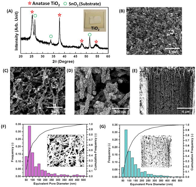
Figure 3.
(A) XRD analysis of the annealed virus-templated anatase TiO2 photoanodes. SEM images of the virus-templated TiO2 photoanodes in (B)-(D) top-down view and (E) cross-section view. The pore size distributions calculated by the SEM images in (F) top-down view and (G) cross-section view, respectively.

