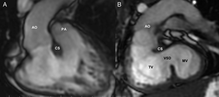
Figure 1:
Preoperative cardiac MRI [Patient 1] (SDD, DORV and VSD): (A) 2D-SSFP (FIESTA) image (coronal view) showing the origin of both great arteries from the right ventricle, with aorta (Ao) to the right of the pulmonary artery (PA). (B) 2D-SSFP (FIESTA) image from an oblique stack demonstrating the left ventricle (LV)-to-aorta route across the ventricular septal defect (VSD). The conoventricular VSD is seen to extend significantly into the inlet region. Tricuspid valve (TV) and the subaortic conal septum (CS) pose potential barriers to the intraventricular tunnelling. Resection of the conal septum and multiple patch tunnelling allowed an unobstructed LV-to-aorta routing for biventricular correction. Ao: aorta; CS: subaortic conal septum; LV: left ventricle; MV: mitral valve; TV: tricuspid valve.

