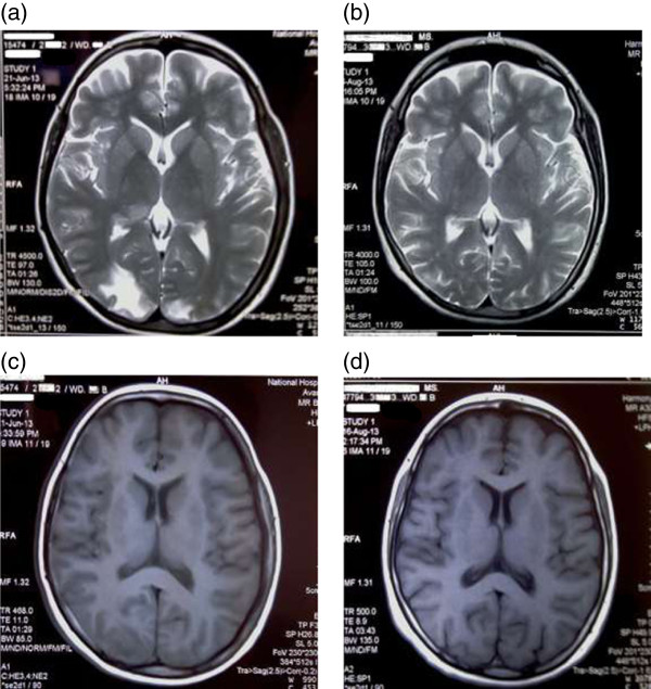
Figure 4.
Preliminary and reassessment MRI brain: (a), (c) are preliminary MRI images taken on presentation. (a) - T2 weighted shows high signal intensity and (c) – T1 weighted shows low signal intensity involving the posterior occipital areas with cortical and subcortical regions. (b), (d) are MRI images taken on reassessment showing improvement.

